
AKIS Imaging
Our current elastography work focuses on the heart, and specifically passive mechanical measures of cardiac stiffness. We estimate the passive mechanical properties by measuring the strain in the ventricles during atrial contraction. The ventricles are usually relaxed during atrial contraction so expansion in the ventricles provides an approximate estimate of passive ventricular stiffness. We're developing this method as an ultrasonic tool to differentiate acute and chronic myocardial infarct.
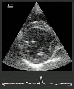
The movie above shows the expansion from the active filling caused by atrial contraction, which is sometimes referred to as the atrial kick. The atrial kick is a small quick expansion and occurs just after the p-wave, which is the first feature in the ECG trace below the ultrasound movie. The movie is slowed down by about a factor of 6.
To give a more complete picture, and to contrast most existing cardiac elastography approaches a cardiac timing diagram is shown below.
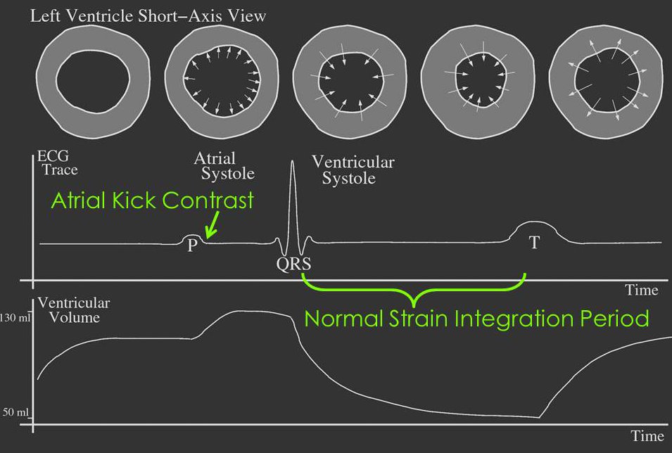
The figure shows a short-axis cartoon of the left ventricle as well as the direction of ventricular wall motion during the different phases. Below the ventricle diagram is the electrocardiogram of the heart, and below these two diagrams is a left ventricle volume chart. The volume chart shows that just after the p-wave a rapid but small increase in volume occurs. The increase in volume induces a small (usually) passive strain in the ventricles.
We estimate the motion in 3D and then we display the axial strain magnitude. (More information on the motion estimation can be seen under Bayesian speckle tracking.)
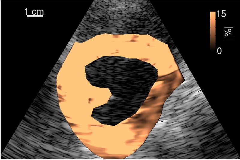
The axial strain magnitude is estimted from each of the axial strains (axial, lateral, and elevational) estimated from the AKIS displacements. The image at right shows an AKIS image of a chronic myocardial infarct. The dark copper shaded pixels correlate to stiff scar tissue and the bright copper pixels correlate to relatively softer healthy myocardium.
Brett Byram

Office: SC 5905
Phone: (615) 343-2327
b.byram _at_ vanderbilt.edu